Posted on

Titanium vs. Stainless Steel Screws in CT and MRI: What Artifact Size Means

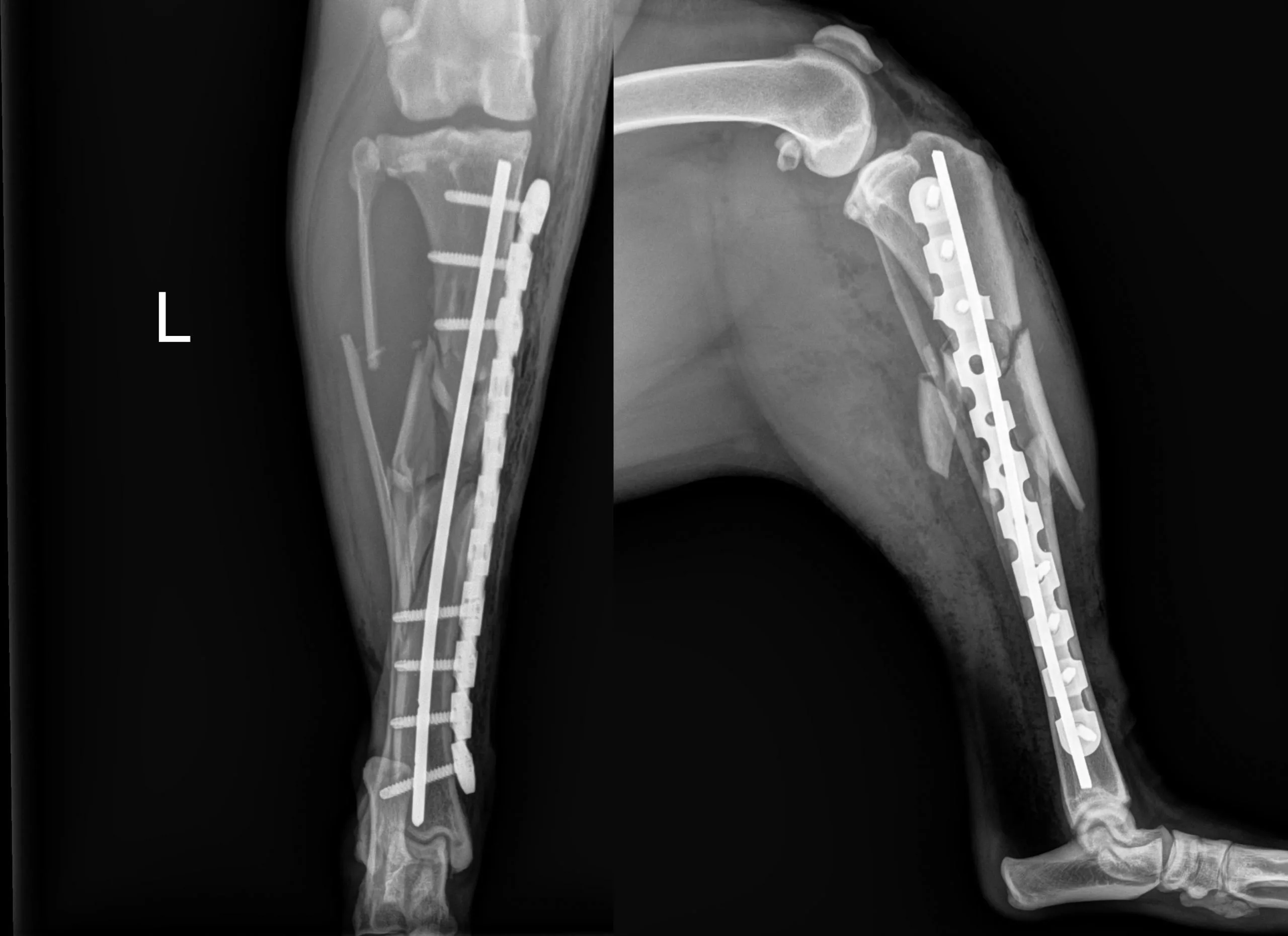
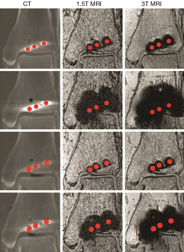
CT, 1.5T MRI and 3T MRI comparison of metal artifacts around orthopedic screws

Published in: Quantitative Imaging in Medicine and Surgery, Volume 4, Issue 3, pages 163–172 · June 2014 · Original research.

Postoperative CT and MRI can help assess bone and joint alignment, but metal implants distort the surrounding image. This cadaveric study compared titanium-alloy and stainless-steel screws to quantify how implant material, screw design and imaging modality change the size of that distortion.

What the study found

CT produced the smallest artifacts overall. For standard screws, the reported artifact radius was approximately 2.0 mm for titanium alloy and 2.6 mm for stainless steel. The difference became much larger in MRI: at 1.5T, titanium and stainless-steel screws produced artifacts of about 3.7 mm and 10.9 mm; at 3T, the corresponding values were about 4.4 mm and 15.3 mm.

Cannulated titanium screws also produced smaller artifacts than cannulated stainless-steel screws. The authors concluded that 1.5T MRI may be a useful non-ionizing option near titanium hardware when adequate distance and image quality are available, while stainless-steel artifacts can obscure a substantially larger region.

Why this matters for veterinary orthopedics

  • Follow-up imaging: implant material can influence how much anatomy remains visible around a fixation construct.
  • Modality selection: CT minimized artifact in this experiment, while field strength strongly affected MRI results.
  • Preoperative planning: anticipated postoperative imaging needs may be one factor in implant selection, alongside strength, geometry, biology and surgical technique.

Clinical context: The experiment used a human cadaver ankle and specific scanner protocols. The measurements should not be transferred directly to every veterinary patient, implant system or imaging sequence.

Read the source

Radzi S, Cowin G, Robinson M, et al. “Metal artifacts from titanium and steel screws in CT, 1.5T and 3T MR images of the tibial pilon: a quantitative assessment in 3D.” Quant Imaging Med Surg. 2014;4(3):163–172. DOI: 10.3978/j.issn.2223-4292.2014.03.06.
Posted on

Stress Modulation in Fracture Fixation: Building a Construct That Can Heal

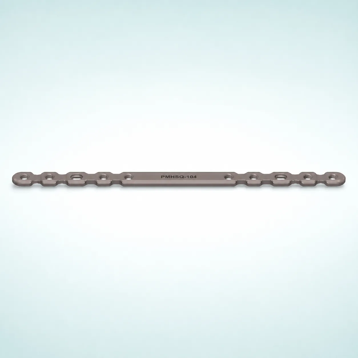
Intact OsteoCertus PMHSQ-104 high-strength medium locking plate

Published in: Journal of the American Academy of Orthopaedic Surgeons (JAAOS), Volume 24, Issue 10, pages 711–719 · October 2016 · Review article.

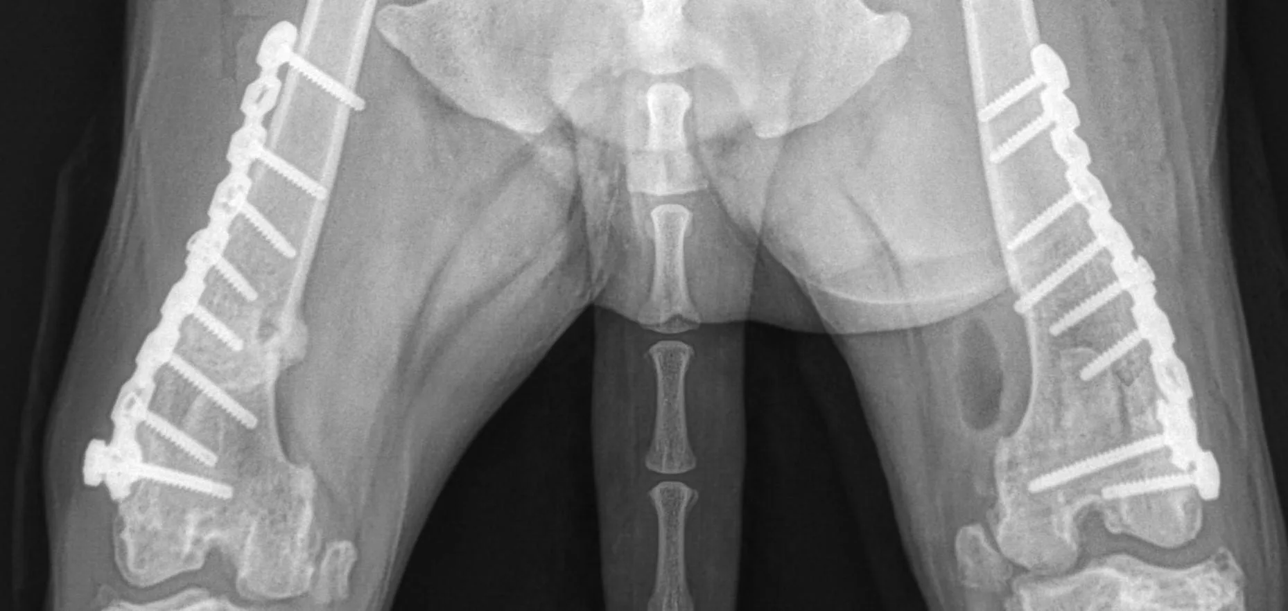
Fracture fixation is not simply a choice between a “strong” and a “weak” construct. The goal is to create enough stability to control the fracture while preserving the small, controlled motion that supports callus formation during secondary bone healing.

Why construct stiffness matters

An overly rigid bridge construct may limit beneficial interfragmentary motion and contribute to asymmetric callus formation. A construct that is too flexible can permit excessive strain, delayed healing, malalignment or fatigue failure. Stress modulation is the deliberate adjustment of implant and technique variables to stay between those extremes.

Variables the surgeon can control

  • Plate span and working length: longer plates and thoughtful screw spacing can distribute load more gradually.
  • Screw density: filling every available hole is not always the most biologically favorable strategy in bridge plating.
  • Material and geometry: titanium is less stiff than stainless steel, but plate thickness, shape and hole design also strongly affect the final construct.
  • Locking strategy: locking, nonlocking and hybrid configurations transfer load differently and should be selected for the fracture pattern and bone quality.

Relevance for veterinary orthopedic surgery

The review was written for human orthopedics, but its biomechanical framework is useful when planning veterinary fracture fixation. Patient size, activity, fracture biology, bone quality and postoperative management must all be considered; material choice alone does not determine clinical success.

Clinical context: This educational summary does not replace case-specific surgical judgment or manufacturer instructions.

Read the source

Beltran MJ, Collinge CA, Gardner MJ. “Stress Modulation of Fracture Fixation Implants.” J Am Acad Orthop Surg. 2016;24(10):711–719. DOI: 10.5435/JAAOS-D-15-00175.

Posted on

Biomechanical Analysis of Rabbit Radius and Humerus Fixation Techniques

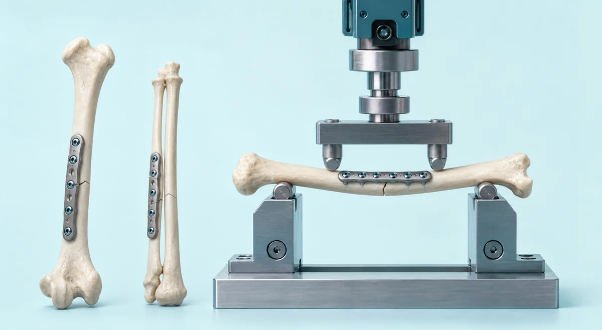
Rabbit long-bone fixation constructs in a four-point biomechanical bending test

Published in: The Canadian Veterinary Journal, Volume 63, Issue 5, pages 521–527 · May 2022 · Peer-reviewed original research.

Choosing a fixation method for rabbit long-bone fractures can be challenging because the radius and humerus are small, loads vary by anatomic site and published comparative data are limited. A study from Red Bank Veterinary Hospital evaluated three fixation strategies in ex vivo rabbit radius and humerus models.

Study design

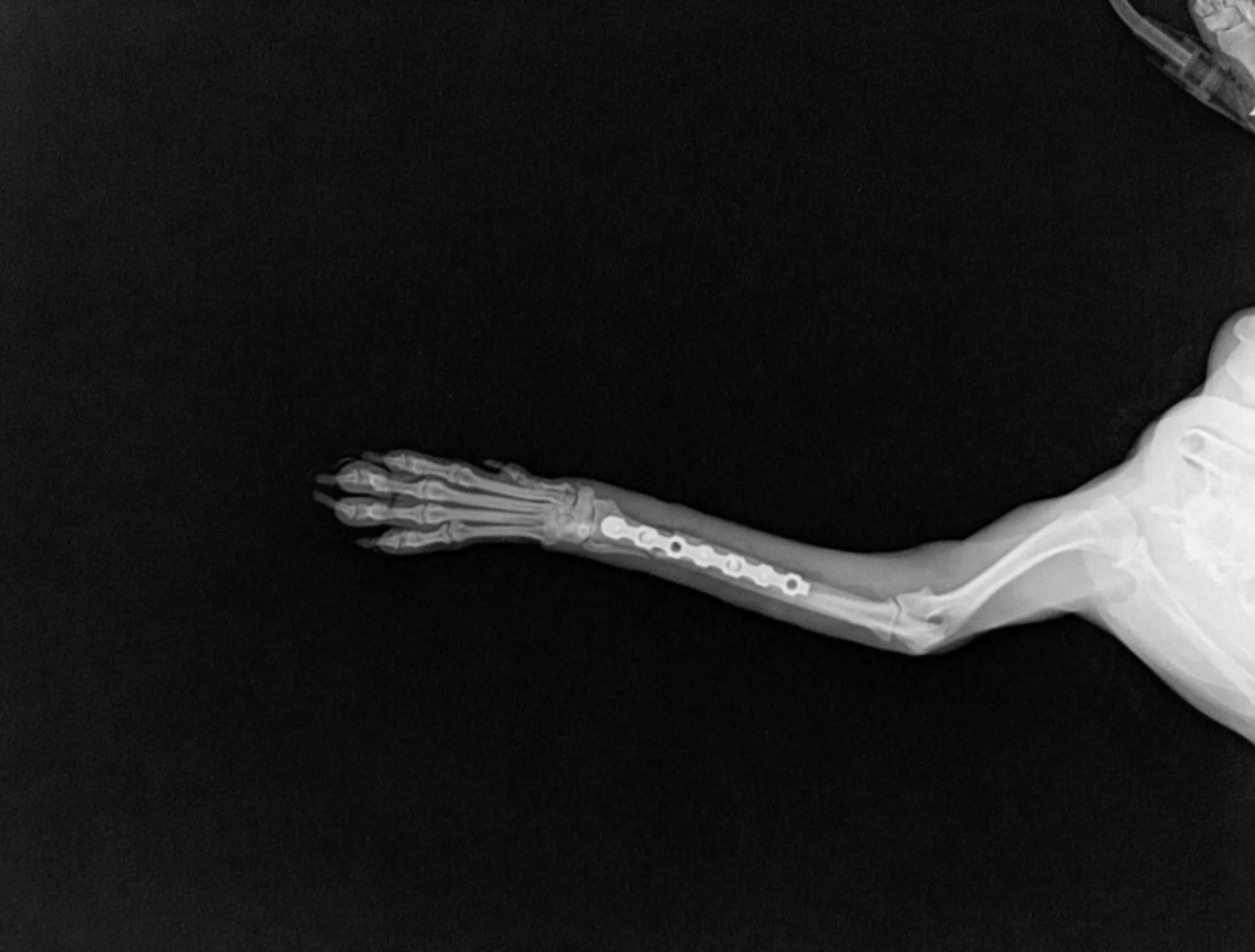
Meghan L. Davolt, Ella Davis, Brynn McCleery and Garrett Davis compared a locking plate construct, a veterinary cuttable plate construct and an external skeletal fixation construct using four-point bending. The locking plate group used a titanium OsteoCertus cuttable locking plate.

What the testing showed

  • In the humerus model, the locking plate construct was the strongest of the three tested methods and most closely approximated the intact bone response.
  • In the radius/ulna model, both plate constructs were stiffer than the external skeletal fixator construct.
  • The radius/ulna testing did not identify a significant difference in strength between the locking plate and veterinary cuttable plate constructs.
  • The results reinforce that implant performance depends on the bone, construct and loading mode; bench testing should inform—not replace—clinical judgment.

Why it matters

The work provides useful comparative mechanical data for surgeons planning rabbit fracture repair. It also illustrates how locking plate technology may contribute to construct stability in small-bone fixation, while patient-specific anatomy, soft tissues and postoperative management remain essential clinical considerations.

Citation: Davolt ML, Davis E, McCleery B, Davis G. Biomechanical analysis of 3 fixation techniques in rabbit radius and humerus bones. Canadian Veterinary Journal. 2022;63(5):521–527. PMID: 35502249.

Read the peer-reviewed article on PubMed Central.

This is an original educational summary. It does not reproduce the journal article and is not a substitute for patient-specific surgical planning.

Posted on

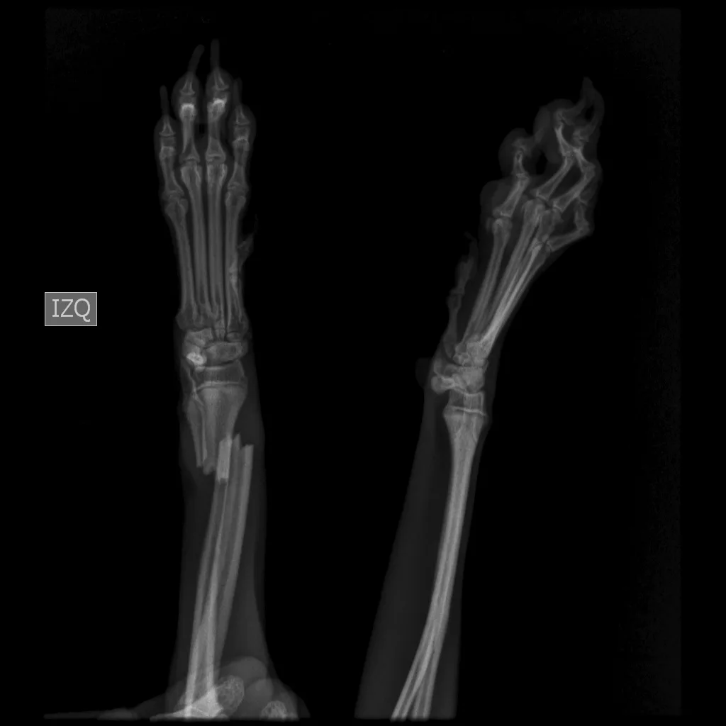
Common Fractures of the Radius and Ulna: Surgical Considerations

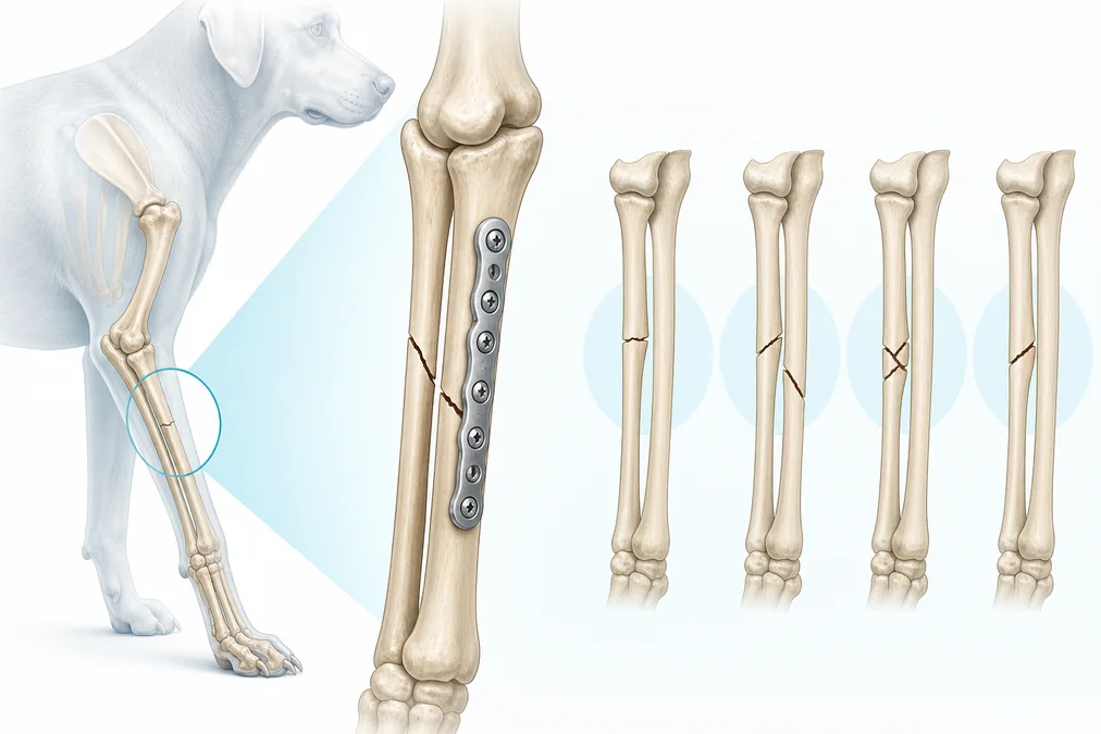
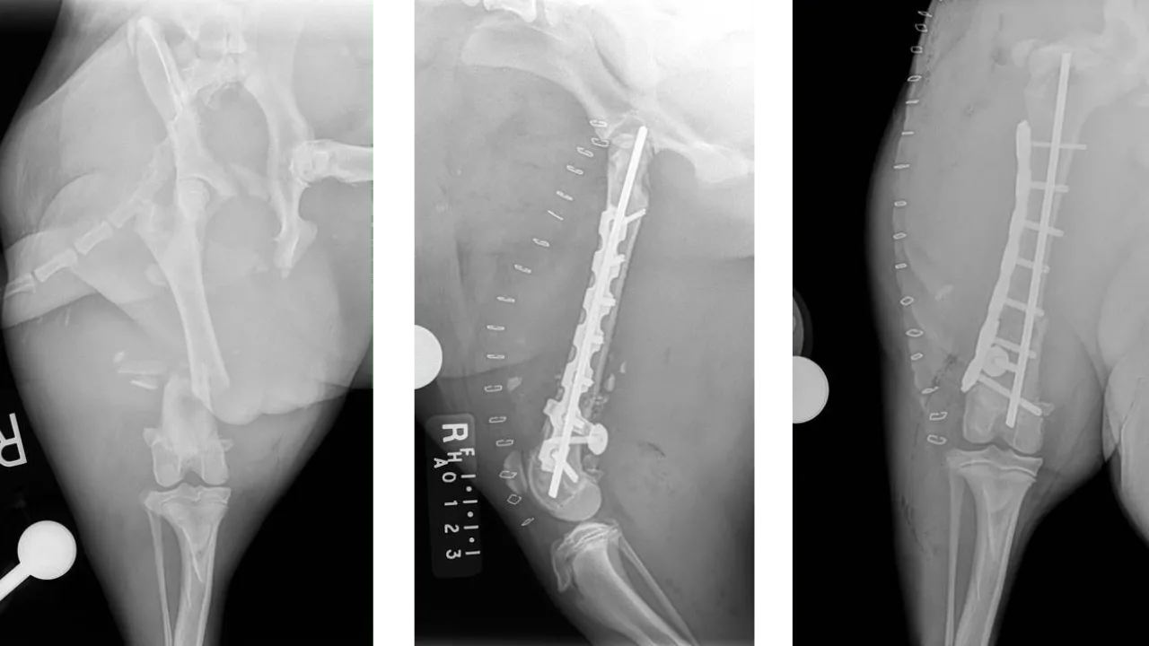
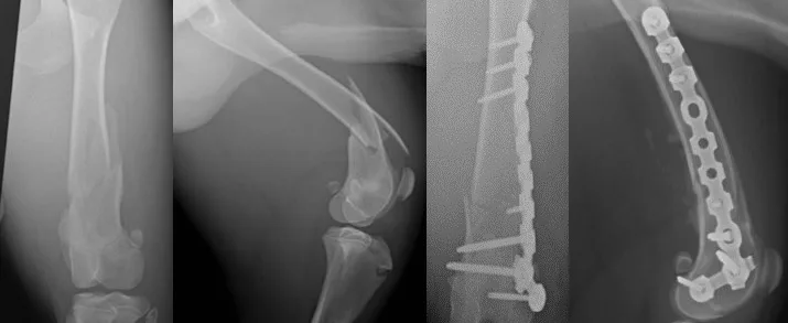
Canine radius and ulna fracture patterns with locking plate repair illustration

Published in: REMEVET, 6th Edition · Presented for CVDL Florida 2026 · Clinical education.

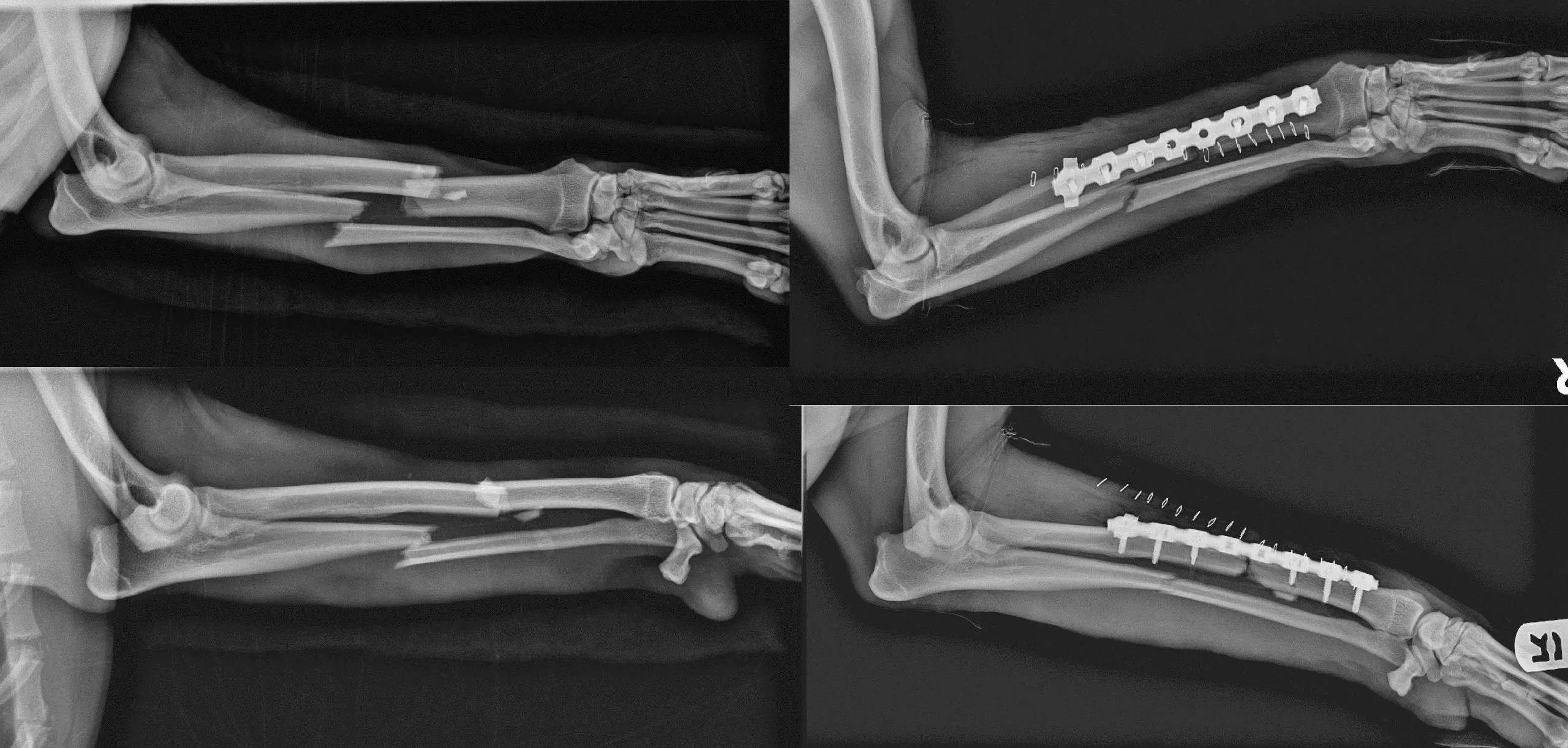
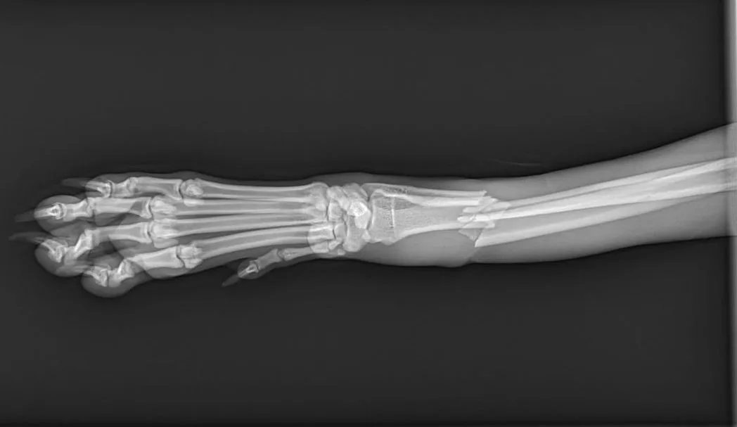
Forelimb fractures demand a fixation plan that respects both mechanics and biology. In this clinical review, Jeff Mayo, MS, DVM, DABVP (C/F), FF-DABVP-CFO, MANZCVS discusses common fracture patterns of the radius and ulna, with related considerations for distal humeral injuries.

Practical points for case planning

  • Define the fracture personality: location, configuration, comminution, soft-tissue condition and patient activity all influence the construct.
  • Preserve biology: limited exposure and minimally invasive plate osteosynthesis may help protect vascularity when the fracture pattern and reduction strategy permit.
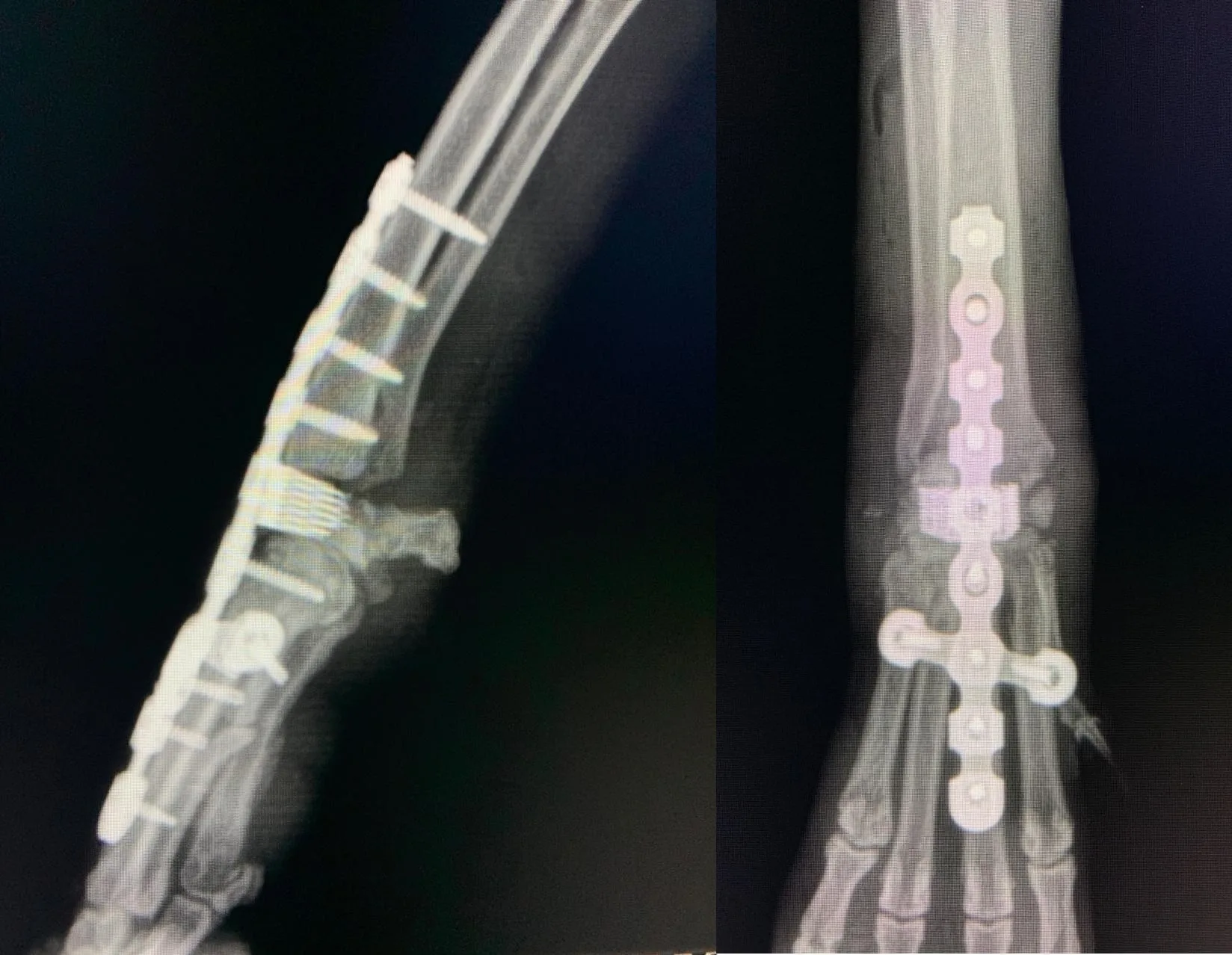
  • Match the implant to the anatomy: plate shape, screw trajectory and available bone stock are especially important in the distal radius and ulna.
  • Plan the complete construct: implant length, working length, screw distribution and postoperative loading should be considered together.
  • Monitor recovery: serial clinical and radiographic evaluation guides activity progression and any decision about later implant removal.

From principles to patient-specific decisions

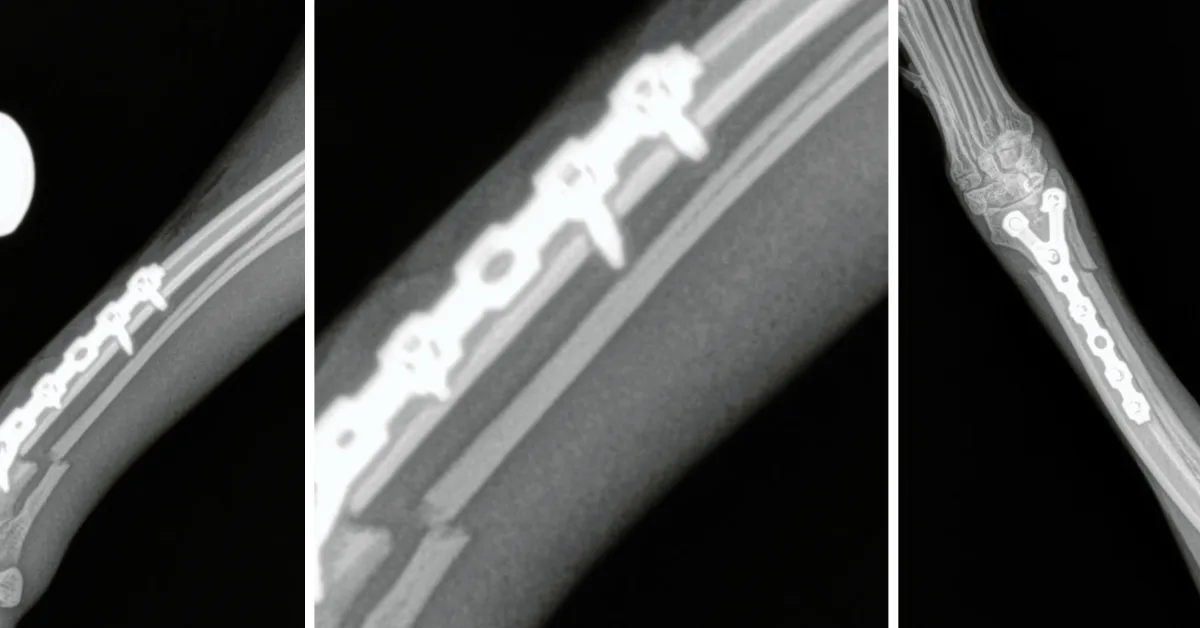
The article highlights plating options—including anatomically adapted and Y-shaped constructs—while emphasizing that no single technique is appropriate for every patient. Careful preoperative planning and atraumatic surgical execution remain central to a successful outcome.

Read the complete REMEVET CVDL Florida 2026 edition.

This summary is intended for veterinary professional education and does not replace patient-specific diagnosis or surgical judgment.

Posted on

Titanium 6Al-4V ELI vs. Stainless Steel 316LVM for Biomedical Implants

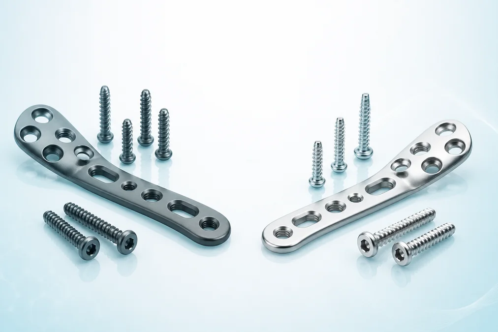
Titanium and stainless steel veterinary orthopedic plates and screws compared on a clinical background

Published in: REMEVET, 6th Edition · Presented for CVDL Florida 2026 · Technical review.

Material selection influences the mechanical behavior, handling and long-term performance of an orthopedic implant. This practical review compares two widely used implant alloys—medical-grade titanium 6Al-4V ELI and stainless steel 316LVM—and explains where their different properties matter in veterinary fracture fixation.

Key differences for orthopedic applications

  • Weight: titanium has a substantially lower density, allowing lighter implants of comparable geometry.
  • Strength-to-weight ratio: titanium combines high mechanical strength with low mass.
  • Elastic modulus: titanium is less stiff than stainless steel and more closely approaches the behavior of cortical bone, an important consideration in load sharing.
  • Corrosion resistance and biocompatibility: the stable oxide layer on titanium supports excellent resistance in physiological environments.
  • Manufacturing and cost: stainless steel remains economical, familiar and comparatively easy to machine.

Clinical interpretation

Neither alloy replaces sound case planning. Implant geometry, working length, screw distribution, patient weight, activity, bone quality and the biological environment all remain essential. The material should be selected as one component of the complete fixation strategy.

This educational article was prepared by Juan Sebastian Silva, Javier Castañeda, Cesar Silva and David Silva and presented in the sixth edition of the REMEVET CVDL Florida magazine.

Read the complete REMEVET CVDL Florida 2026 edition.

For veterinary professional education. Product choice and surgical planning should be based on the individual patient and the treating surgeon’s clinical judgment.

Posted on

Photobiomodulation and Tibial Fracture Healing in a Puppy

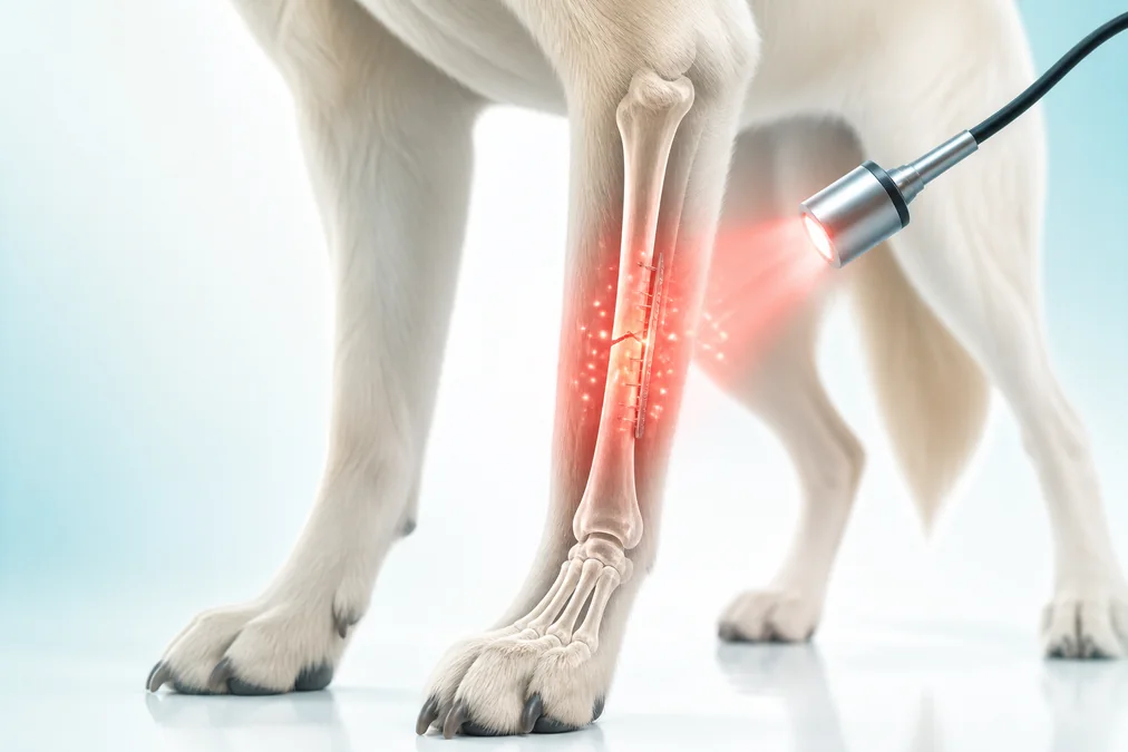
Photobiomodulation applied to a healing canine tibial fracture illustration

Published in: Veterinary Medicine and Science, Volume 9, Issue 2, pages 591–599 · March 2023 · Peer-reviewed case report.

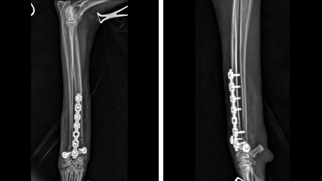
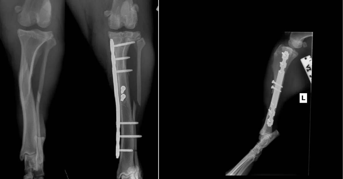
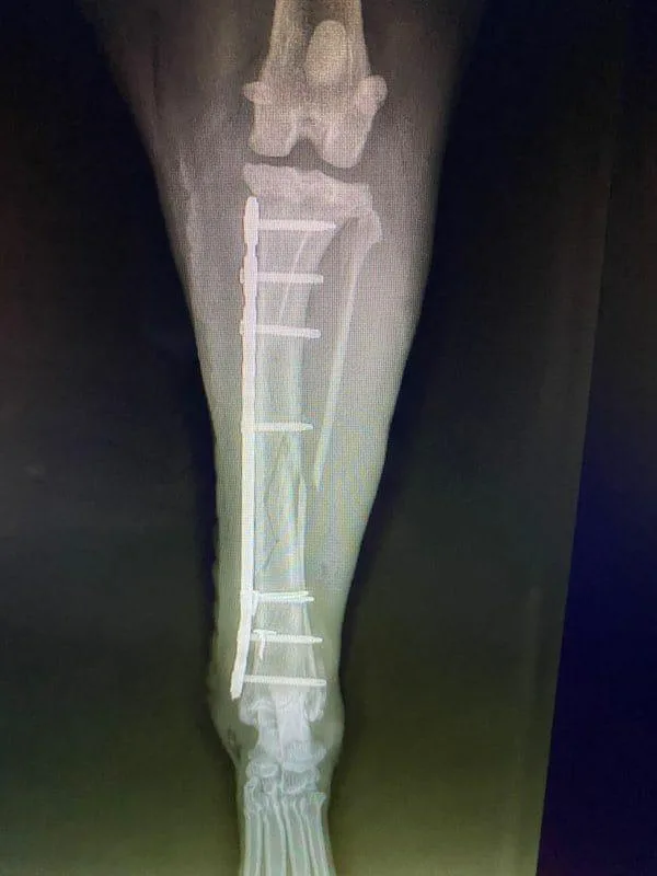
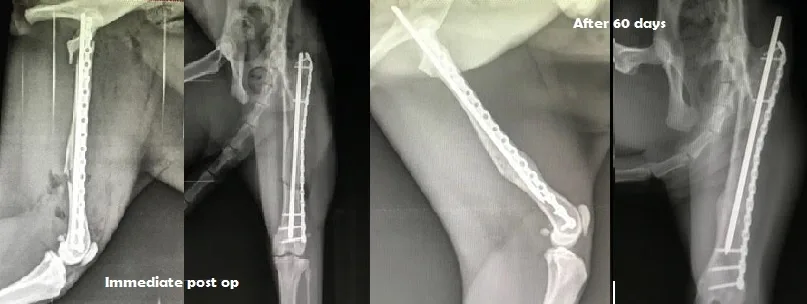
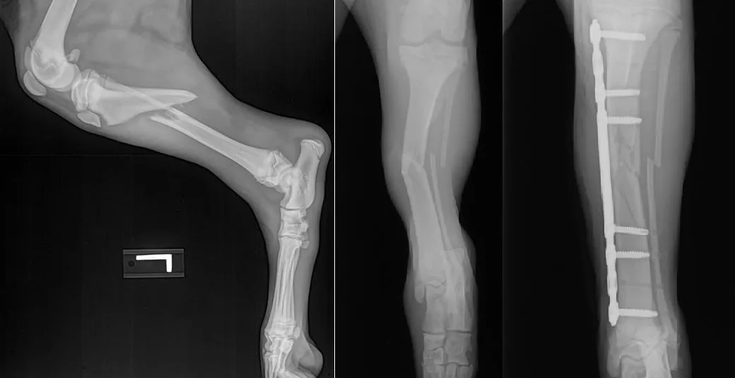
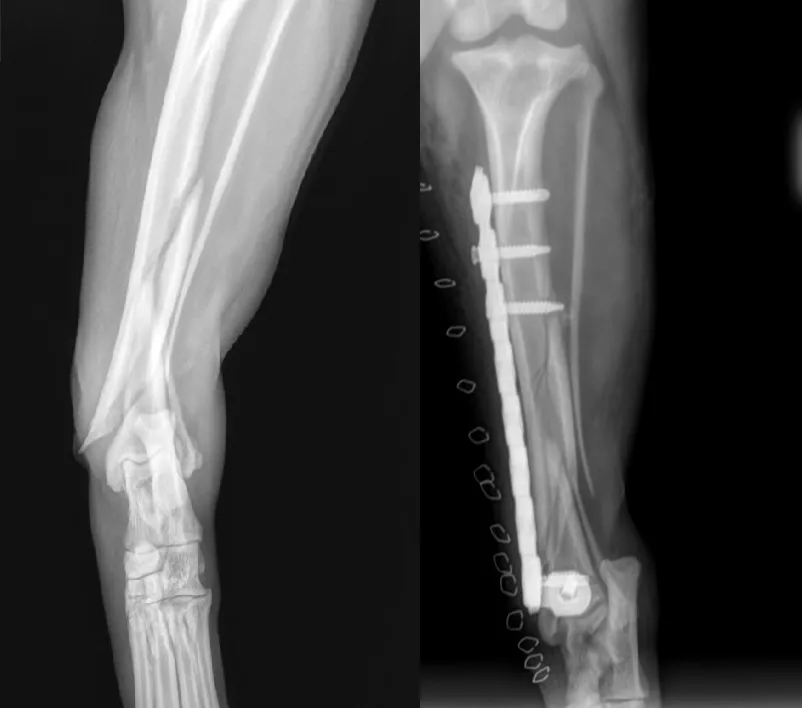
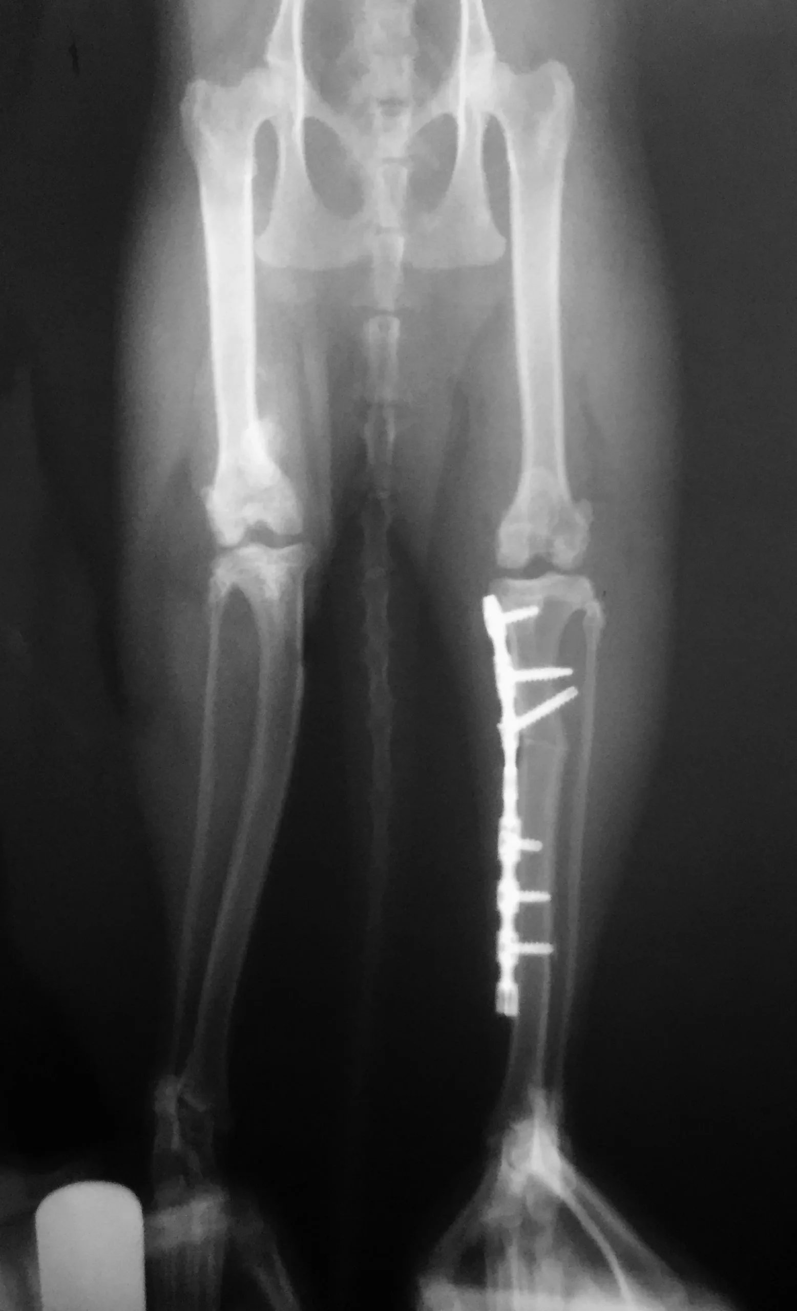
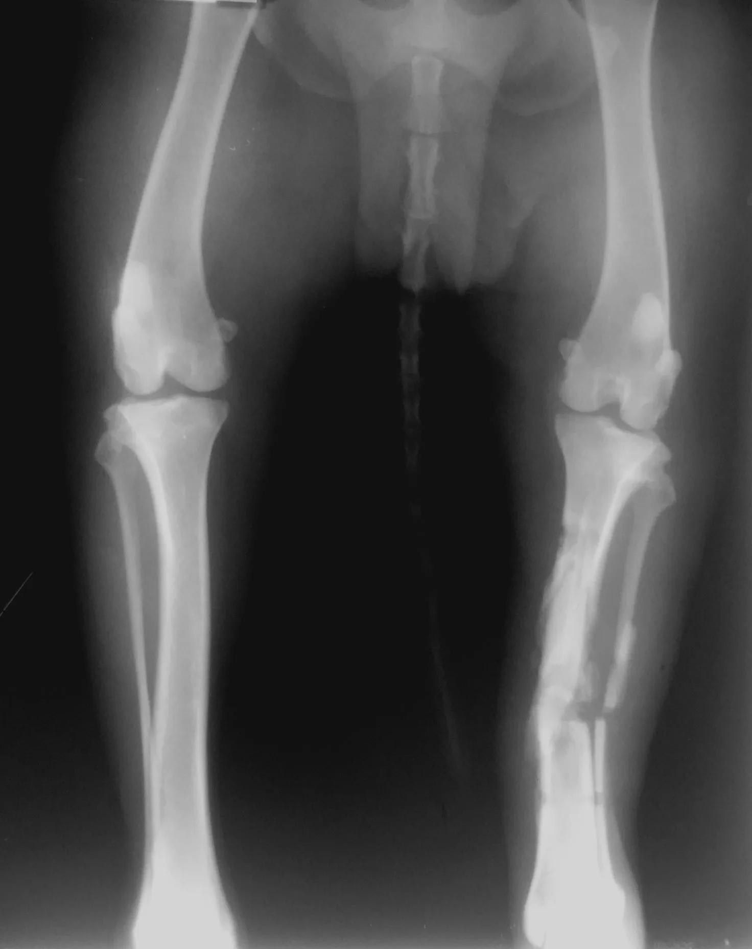
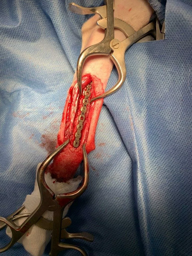
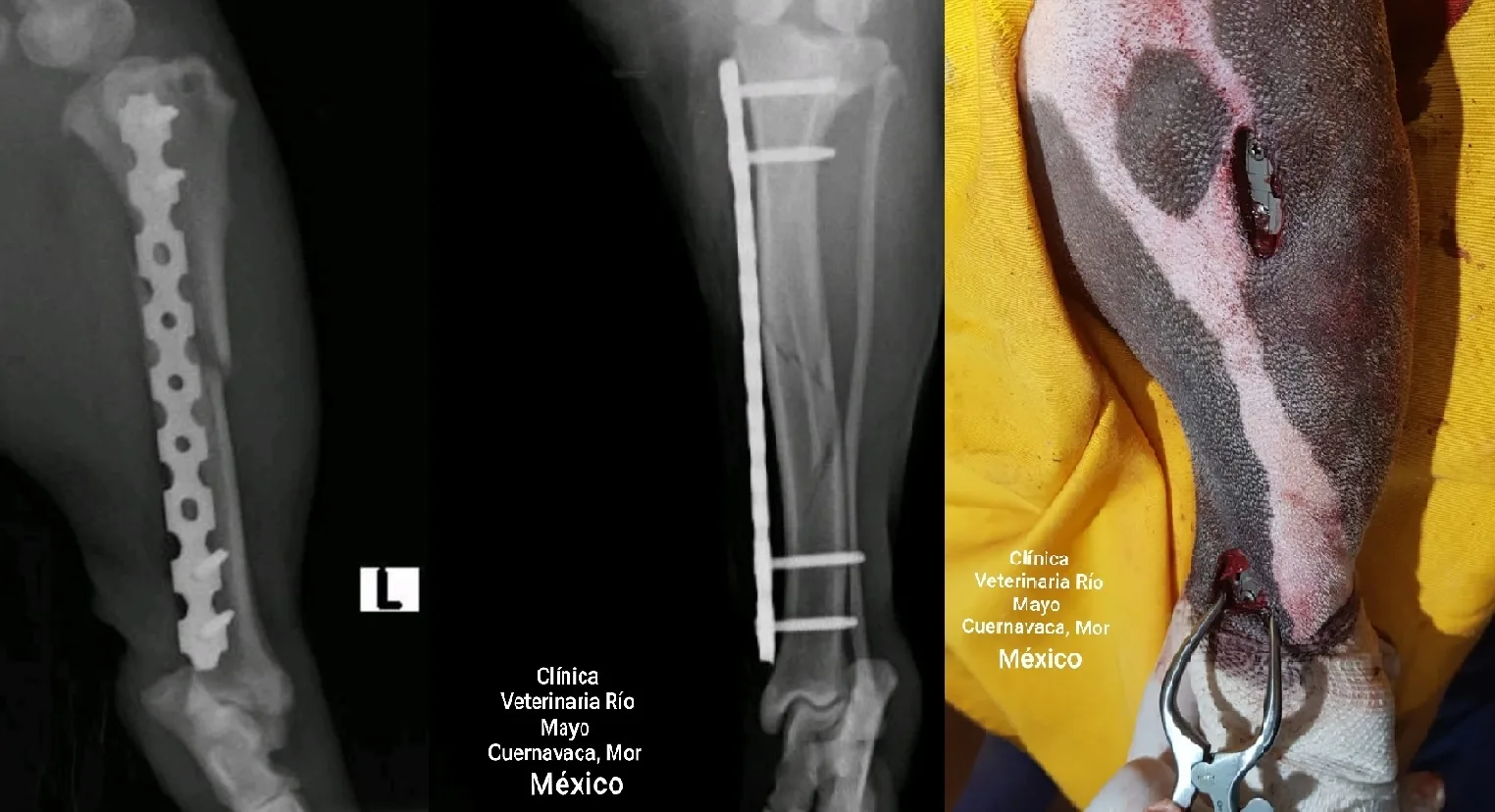
A 10-week-old, 8.9 kg Xoloitzcuintle puppy presented with a complete transverse fracture of the right proximal tibia. The case was managed with minimally invasive plate osteosynthesis (MIPO) using an OsteoCertus titanium locking plate, followed by adjunct photobiomodulation therapy combined with a static magnetic field (PBMT-sMF).

Treatment and follow-up

  • The locking plate was applied using a biologically oriented, minimally invasive approach.
  • PBMT-sMF was delivered twice daily for 21 days at sites proximal to, distal to and over the fracture zone.
  • Radiographs at day 18 showed advancing bone healing with the fracture line still visible.
  • At day 55, the report documented enlarged callus, remodeling and radiographic and clinical union.

Clinical perspective

The report illustrates the combination of stable fixation, biological fracture management and a non-invasive adjunct therapy. Because this is a single case without a control group, it cannot establish that PBMT-sMF caused the observed healing response. Controlled veterinary studies are needed to define indications, dosing and comparative benefit.

Citation: Asteinza Castro IM, Amador Morga A, Johnson DS. Photobiomodulation therapy combined with static magnetic field in tibial fracture healing of a dog: a case report. Veterinary Medicine and Science. 2023;9(2):591–599. PMID: 36639946.
Read the open-access article on PubMed Central.
This summary is intended for veterinary professional education and does not replace patient-specific diagnosis, treatment planning or clinical judgment.