
Published in: Quantitative Imaging in Medicine and Surgery, Volume 4, Issue 3, pages 163–172 · June 2014 · Original research.
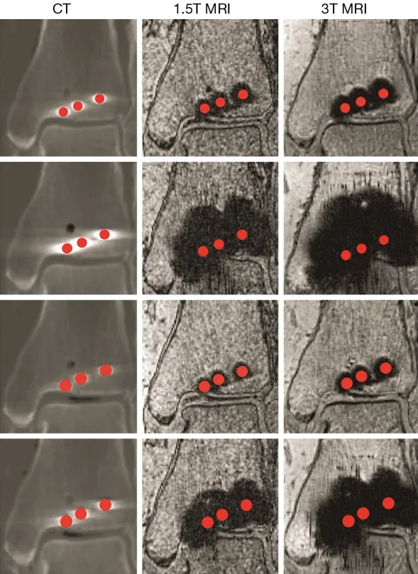
Postoperative CT and MRI can help assess bone and joint alignment, but metal implants distort the surrounding image. This cadaveric study compared titanium-alloy and stainless-steel screws to quantify how implant material, screw design and imaging modality change the size of that distortion.
What the study found
CT produced the smallest artifacts overall. For standard screws, the reported artifact radius was approximately 2.0 mm for titanium alloy and 2.6 mm for stainless steel. The difference became much larger in MRI: at 1.5T, titanium and stainless-steel screws produced artifacts of about 3.7 mm and 10.9 mm; at 3T, the corresponding values were about 4.4 mm and 15.3 mm.
Cannulated titanium screws also produced smaller artifacts than cannulated stainless-steel screws. The authors concluded that 1.5T MRI may be a useful non-ionizing option near titanium hardware when adequate distance and image quality are available, while stainless-steel artifacts can obscure a substantially larger region.
Why this matters for veterinary orthopedics
- Follow-up imaging: implant material can influence how much anatomy remains visible around a fixation construct.
- Modality selection: CT minimized artifact in this experiment, while field strength strongly affected MRI results.
- Preoperative planning: anticipated postoperative imaging needs may be one factor in implant selection, alongside strength, geometry, biology and surgical technique.
Clinical context: The experiment used a human cadaver ankle and specific scanner protocols. The measurements should not be transferred directly to every veterinary patient, implant system or imaging sequence.
Read the source
Radzi S, Cowin G, Robinson M, et al. “Metal artifacts from titanium and steel screws in CT, 1.5T and 3T MR images of the tibial pilon: a quantitative assessment in 3D.” Quant Imaging Med Surg. 2014;4(3):163–172. DOI: 10.3978/j.issn.2223-4292.2014.03.06.