
Published in: REMEVET, 6th Edition · Presented for CVDL Florida 2026 · Clinical education.
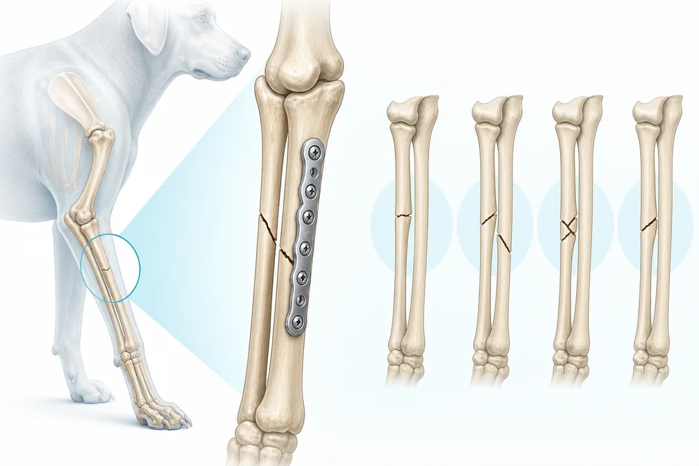
Forelimb fractures demand a fixation plan that respects both mechanics and biology. In this clinical review, Jeff Mayo, MS, DVM, DABVP (C/F), FF-DABVP-CFO, MANZCVS discusses common fracture patterns of the radius and ulna, with related considerations for distal humeral injuries.
Practical points for case planning
- Define the fracture personality: location, configuration, comminution, soft-tissue condition and patient activity all influence the construct.
- Preserve biology: limited exposure and minimally invasive plate osteosynthesis may help protect vascularity when the fracture pattern and reduction strategy permit.
- Match the implant to the anatomy: plate shape, screw trajectory and available bone stock are especially important in the distal radius and ulna.
- Plan the complete construct: implant length, working length, screw distribution and postoperative loading should be considered together.
- Monitor recovery: serial clinical and radiographic evaluation guides activity progression and any decision about later implant removal.
From principles to patient-specific decisions
The article highlights plating options—including anatomically adapted and Y-shaped constructs—while emphasizing that no single technique is appropriate for every patient. Careful preoperative planning and atraumatic surgical execution remain central to a successful outcome.
Read the complete REMEVET CVDL Florida 2026 edition.
This summary is intended for veterinary professional education and does not replace patient-specific diagnosis or surgical judgment.